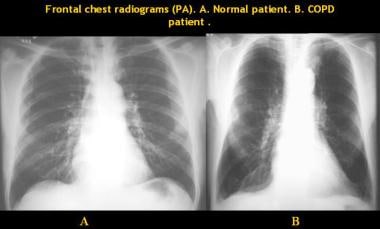
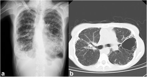
Bilateral Emphysematous Lung Fields

Chronic Obstructive Lung Disease Copd Emphysema

Chest X Ray Showing Predominant Emphysematous Changes Along With A Download Scientific Diagram
Emphysema Imaging Practice Essentials Radiography Computed Tomography
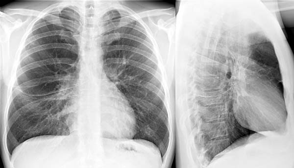
Teaching File Radiologic Anatomy Of The Lung

Bilateral Pulmonary Hyperaeration Metabolic Disorders

Lung Hyperinflation Radiology Reference Article Radiopaedia Org

A collapsed lung is an uncommon but serious condition that can be life threatening for people in advanced stages of emphysema.
Bilateral emphysematous lung fields.

Chronic Obstructive Diseases Of The Lung Part 3
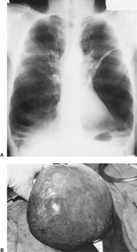
Bullous And Bleb Diseases Of The Lung Thoracic Key

Persistent Pulmonary Interstitial Emphysema In A Case Of Langerhans Cell Histiocytosis

Silicosis In The Form Of Progressive Massive Fibrosis A Diagnostic Challenge

Cardiothoracic Imaging Bilateral Emphysema Chronic Obstructive Pulmonary Disease Enlarged Heart Pulmonary
Imaging From A 64 Year Old Man With Cpfe A Hrct Of Bilateral Upper Download Scientific Diagram

Cannabis Bong Smoking Induced Pneumomediastinum And Subcutaneous Emphysema Radiology Case Radiopaedia Org

Pulmonary Interstitial Emphysema Radiology Case Radiopaedia Org
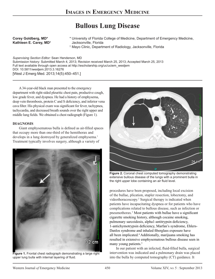
Pdf Bullous Lung Disease

Pin By Kathy I On Radiology Copd Lunges Radiology

Emphysema With Images Copd Lung Disease Lunges

E X20rfsg84elm

Hyperlucent Lung Syndrome Associated With Persistent Foramen Ovale And Bilateral Bullous Emphysema Consultant360
Perioperative Management Of Esophagectomy In A Patient Who Previously Underwent Bilateral Lung Transplantation Springerlink

Pdf Emphysema And Copd In A Young Woman

Regional Ventilation And Perfusion After Lung Transplantation In Patients With Emphysema Nejm
Article Fulle Text

Jaypeedigital Ebook Reader
Https Encrypted Tbn0 Gstatic Com Images Q Tbn 3aand9gcs Frib0buwx1mhxnpsgaz8qstkmod16edyuebeh3g Usqp Cau
Pulmonary Emphysema Radiology Reference Article Radiopaedia Org

Ct Scans Showing Tracheomegaly A Bilateral Upper Lobe Emphysema B Download Scientific Diagram

Subcutaneous Emphysema Radiology Case Radiopaedia Org Radiology Radiology Imaging Subcutaneous Emphysema
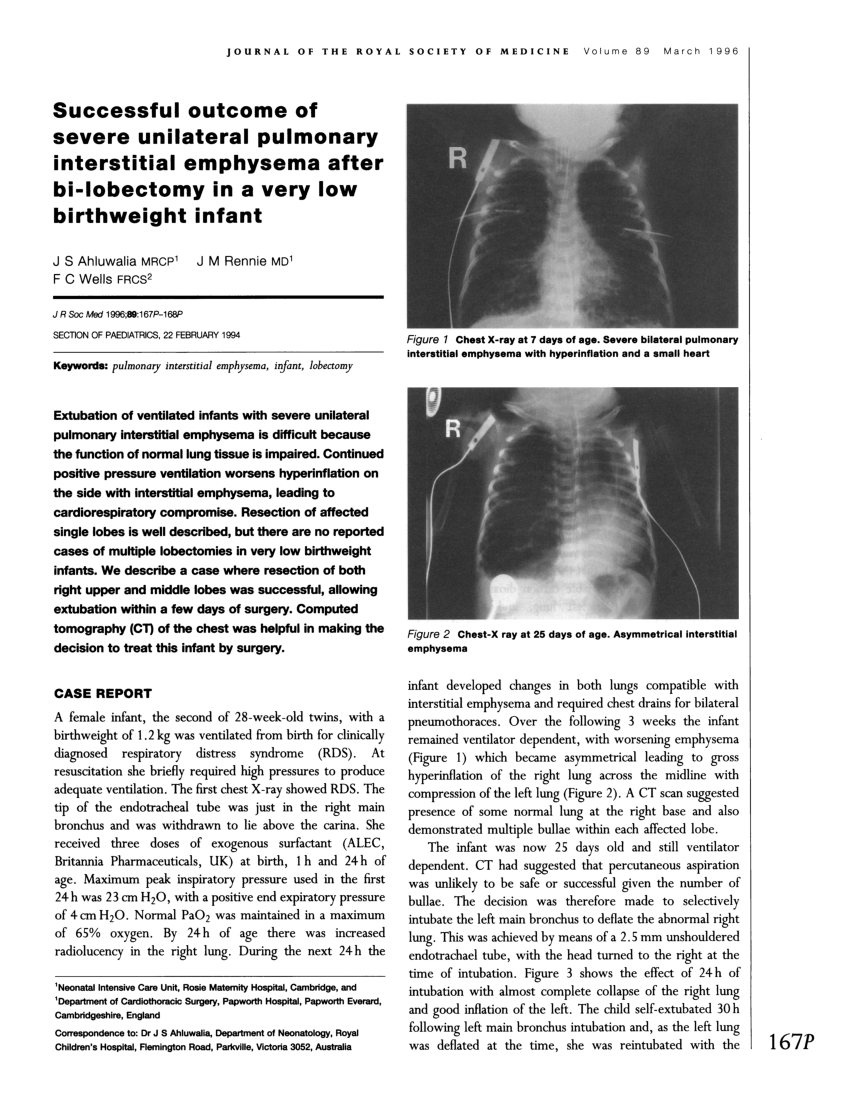
Pdf Successful Outcome Of Severe Unilateral Pulmonary Interstitial Emphysema After Bi Lobectomy In A Very Low Birthweight Infant

Pdf Percutaneous Evacuation Of Diffuse Pulmonary Interstitial Emphysema By Lung Puncture In A Baby With Extremely Low Birth Weight A Case Report
Source : pinterest.com
